Every foot is different, and so is every recovery. These photos show actual patient results at various stages of healing after minimally invasive bunion surgery performed at Fallbrook Podiatry. No two recoveries are identical — your own timeline may vary.
Individual results vary. These photographs are published with patient consent. Photos are not retouched or digitally altered beyond background standardization. All procedures were performed by Dr. Grigoriy N. Patish, DPM.Patient Results
1 Bunion & Hammertoe Correction — Left Foot
Left foot bunionectomy with second toe floating hammertoe correction. This case demonstrates the progressive healing pattern from the day of surgery through 29 days, showing how swelling subsides and toe alignment improves week by week.
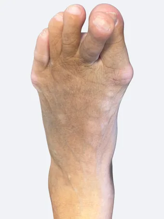 Before
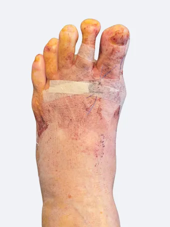 48 hrs
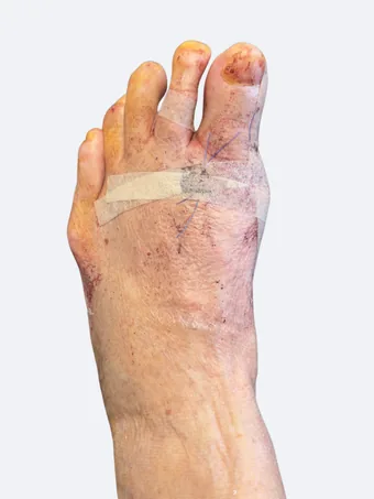 9 days
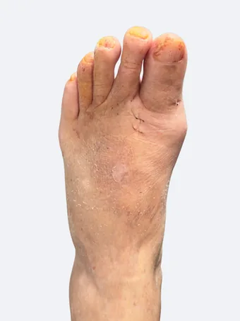 16 days
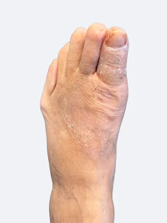 22 days
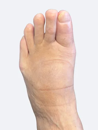 29 days
2 Bunion Correction — Right Foot
Right foot bunion correction in a 28-year-old patient. Day-of-surgery photo shows Betadine (povidone-iodine) antiseptic staining, which is standard sterile preparation — the orange color is not part of the condition. Recovery progression shown through 22 days with suture and strapping visible.
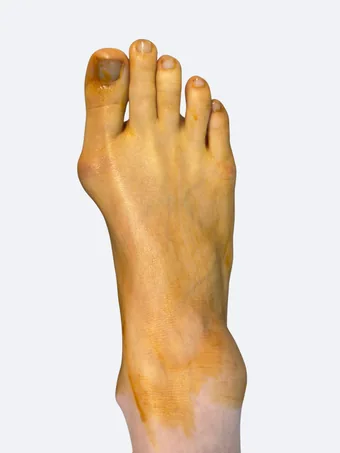 Day of Surgery
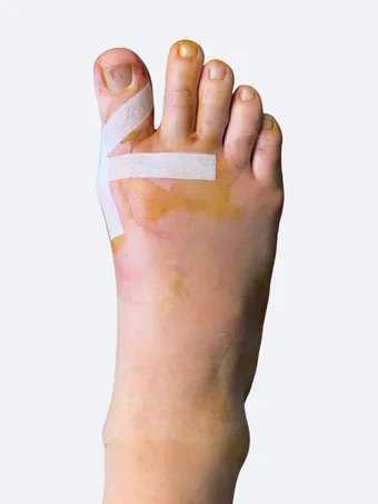 2 days
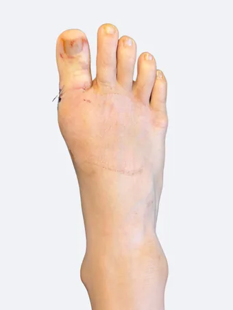 15 days
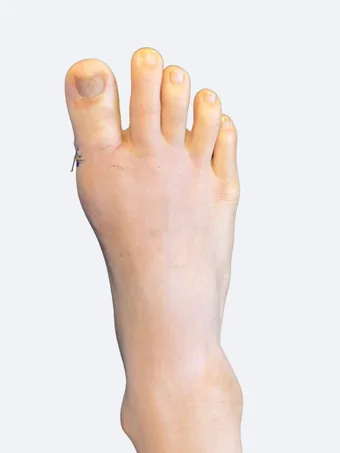 22 days
3 Bunion Correction — Left Foot
Left foot bunion correction in a 20-year-old male patient. Before and early post-operative views: alignment is corrected during surgery, and swelling is expected. Individual results vary.
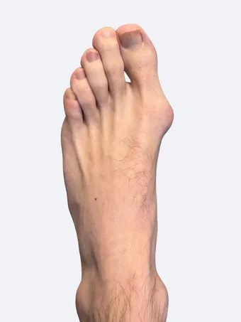 Before
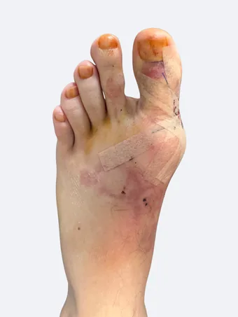 48 hrs
4 Bunion Correction — Left Foot
Left foot bunion correction in a 20-year-old female patient. This case includes the post-operative dressing immediately after surgery and the bandaged foot on day of procedure, providing a complete picture of the surgical and early recovery experience.
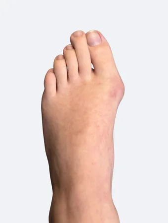 Before
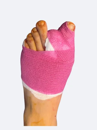 Day of Surgery
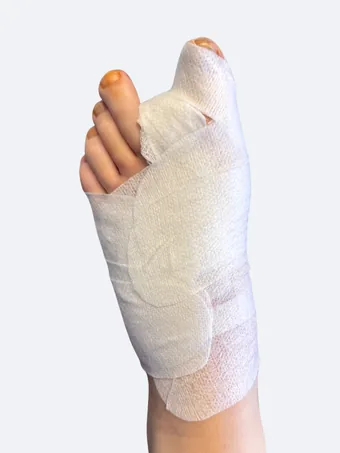 2d dressing
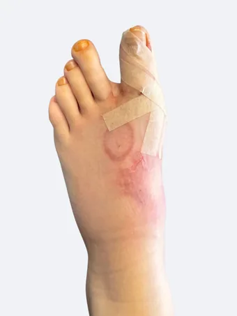 2 days
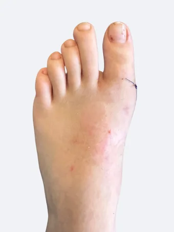 13 days
View X-Ray Comparison
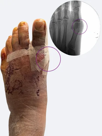
These images combine clinical photography with corresponding X-rays to show both the visible and structural correction achieved through surgery.
5 Bunion Correction with X-Ray — Left Foot
Left foot bunion correction with radiographic comparison. The circled areas highlight the first metatarsophalangeal joint, showing reduction of the intermetatarsal angle and improved joint congruity after surgical correction.
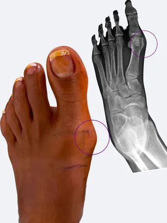 Before
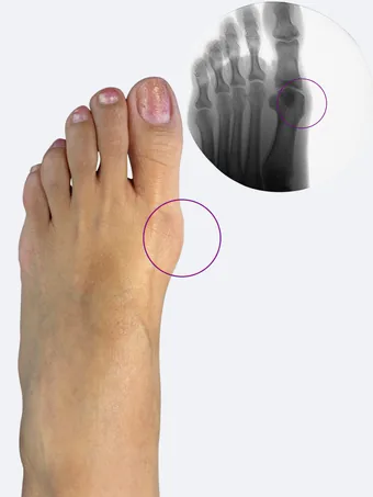 After
After
6 Bunion Correction with X-Ray — Left Foot
Left foot bunion correction with pre-operative and early post-operative X-ray overlay. The early post-operative X-ray shows the corrected metatarsal position, with the expected soft-tissue swelling still present. Individual results vary.
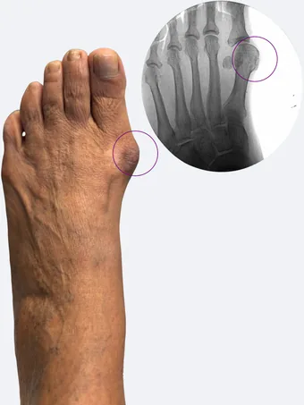 Before
 48 hrs
Considering bunion surgery? Dr. Patish will evaluate your foot, review your X-rays, and give you a straight recommendation about whether minimally invasive correction is right for you.
Schedule a Consultation ← Back to Bunion Surgery Information